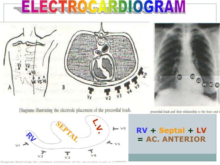
Lead Diagram Septal
Lead Diagram Septal. Learn vocabulary, terms and more with flashcards, games and other study tools. Septal terminals have been identified in most re-gions of the hippocampus, although the most dense projec-. tion is to the subgranular hilar region of the dentate gyrus.

RV infarct, CHB, right sided chest leads.jpg.
Septal terminals have been identified in most re-gions of the hippocampus, although the most dense projec-. tion is to the subgranular hilar region of the dentate gyrus.
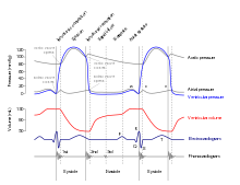
Wiggers diagram , showing a normal ECG curve synchronized ...
Ecg update(basic cardiology)

Field 12 Lead ECG Diagnosis

ECG with atypical atrial flutter with negative flutter ...

Initial EKG, EKG taken at admission notable for q-waves in ...

P/S lead via persistent left superior vena cava placed in ...

Lower atrial septal pacing: A, Fluoroscopic left anterior ...

Chest radiograph of a dual-chamber pacemaker recipient ...

Field 12 Lead ECG Diagnosis
The septal nuclei are subcortical nuclei initially implicated by early ablation and stimulation studies in the regulation of emotional responsiveness such as rage behavior. In particular, it ignores the formation of solid solutions of tin and lead. Lead and Lag can be used in any type of dependency in a network diagram.

0 Response to "Lead Diagram Septal"
Post a Comment