Lead Ekg Limb Diagram
Lead Ekg Limb Diagram. Are the limb leads important in placement for a proper ekg? I've been taught where and so on and so forth, but after reading your blog, I'm betting you'd have a nifty diagram.

The doctor may ask you to hold your breath briefly during the test.
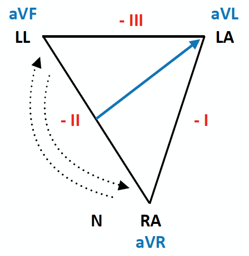
Bipolar Limb leads - Standard Lead I, Lead II and Lead III.

The Monitoring ECG (Clinical Essentials) (Paramedic Care ...

The frontal plane limb leads conventionally represented on ...

ECG Measurement and Analysis

Placement of the 10 electrodes to obtain the 12-lead ...

Patient's 12-lead ECG on admission to the department. Limb ...

The hexaxial reference system. Note that the position of ...

12 Lead ECG Placement | AED Superstore Resource Center
ECG Limb Lead Reversal • LITFL • ECG Library Diagnosis

12 channel ECG & Electrode placement - BPL Medical ...
Unipolar leads (augmented leads and chest leads) have a single positive recording electrode and utilize a combination of the other electrodes to serve as a composite negative electrode. Before exercise commencement, the investigator will perform an EKG at a resting. The six limb leads are called lead I, II, III, aVL, aVR and aVF.

0 Response to "Lead Ekg Limb Diagram"
Post a Comment